

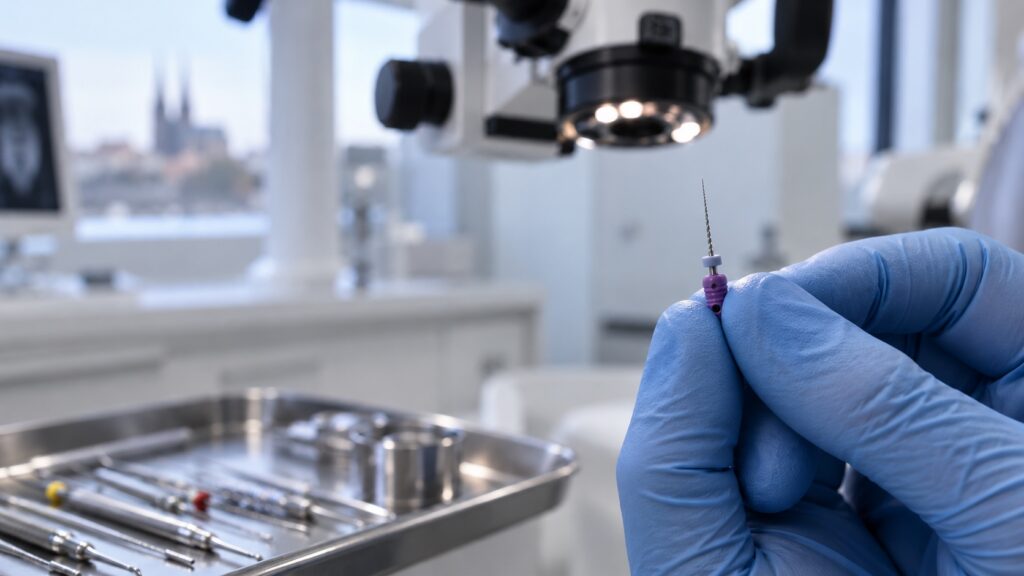
A root canal is not always clearly visible on an X-ray, and conventional instruments cannot always enter it. Hard tissue may gradually form inside, narrowing the space until it closes completely. This is known as a calcified root canal.
Calcification does not automatically mean losing the tooth. Treatment is more demanding because the dentist has to locate and open a space only a fraction of a millimetre wide. This requires an operating microscope and sometimes a 3D scan. The outcome also depends on the root, tooth crown and surrounding bone.
What does it mean when a root canal becomes calcified?
A root canal is a narrow space inside the root containing the dental pulp, commonly called the nerve. During treatment, the dentist locates, cleans and fills it.
Irritation or trauma can cause more hard tissue to form inside. The canal narrows until it may be as thin as a hair or closed along part of its length. It then becomes difficult to see on an X-ray and access with conventional instruments.
Common causes include:
- an impact to the tooth, even many years earlier
- deep decay or long-term irritation of the pulp
- repeated treatment of the same tooth
- prolonged overloading caused by teeth grinding
- increasing age, as root canals may gradually narrow over time
The consequences of an old injury can appear much later, even if the tooth never hurt. By then the canal may be almost closed, with symptoms developing only after the pulp dies.
Why a calcified canal should not be treated without adequate visual control
Without a clear view of the canal, four risks increase:
- The instrument misses the canal, creating a false passage in healthy tissue.
- The root wall is perforated, creating an opening into the surrounding tissues and worsening the prognosis.
- An instrument fractures, blocking access to the rest of the canal.
- The canal is not located, allowing bacteria and inflammation to remain or return.
An operating microscope magnifies and illuminates the inside of the tooth. A 3D scan can show the canal’s route and narrowing. Our article about treatment under a dental microscope explains the benefits; precision is particularly important in calcified canals.
Upper molars may also contain an additional canal that is difficult to find without magnification. If untreated, bacteria can remain and inflammation may return.
How is a tooth with a calcified canal treated?
Treatment is usually more time-consuming than conventional root canal treatment and involves several steps:
- 3D scan. If needed, it shows the canal’s route and narrowing.
- Access. Under a microscope, the dentist approaches the canal according to the tooth’s anatomy.
- Locating the entrance. Hard tissue covering it is removed layer by layer under magnification.
- Opening the canal. Fine instruments advance through it while its length is checked.
- Cleaning and filling. The opened canal is cleaned, disinfected and filled.
- Protecting the tooth. Depending on its strength, the dentist uses a filling or recommends a crown.
Treatment may take two visits. It forms part of endodontics; current costs are in our price list. The scope and price of calcified cases depend on their complexity.
What if the canal cannot be opened?
Sometimes the full length cannot be opened. The next step depends on the tooth and surrounding tissues.
Surgical treatment from the root tip. If the usual route is blocked, the dentist may approach from the root tip, remove the bacterial source and fill the canal from that side.
Monitoring. A symptom-free tooth without inflammation may only need regular follow-up X-rays.
Extraction and replacement. This may be needed if the tooth is cracked, severely weakened or lacks bone support.
If a previously treated tooth develops another problem, root canal retreatment may be considered. Planning depends on what remains inside the canal.
Frequently asked questions
What is a calcified root canal?
It has narrowed or closed because new hard tissue formed inside. It may not show on a standard X-ray, and treatment requires fine instruments, precision and time.
Why has my root canal become calcified?
Common causes are an impact, deep decay, prolonged pulp irritation and repeated treatment. Canals also narrow with age, while trauma may have effects years later.
Can a tooth with calcified root canals be saved?
Often, yes. Treatment usually requires an operating microscope and sometimes a 3D scan. If the canal cannot be opened, root-end surgery may be considered.
Why did another clinic decline to treat it?
Access can be difficult without magnification, and planning may require a 3D scan. Your dentist may refer you to a specialist or suggest another approach to reduce the risk of damage.
Conclusion
A calcified canal does not automatically mean that you will lose the tooth. Treatment does, however, require accurate diagnosis, magnification and sufficient time. Whether the tooth can be saved depends on the patency of the canal as well as the condition of the entire tooth and the surrounding tissues.
Have you been told that your tooth cannot be treated conventionally? Book a consultation at m2stoma in Brno. Based on the examination and, if needed, a 3D scan, we will assess whether it makes sense to attempt treatment of the canal or choose another approach.