Without a microscope, a dentist cannot see the fourth canal in an upper molar. And that is often exactly why, two years on, you think your root canal has failed — even though the dentist did excellent work on everything they could see.
The human eye resolves detail down to around 0.2 mm in ideal conditions, but a lateral canal often measures just 0.1 mm across. What the dentist cannot see, they cannot clean. And if a single untreated canal with bacteria is left behind, the inflammation returns.
That is why the microscope has become indispensable in modern endodontics. A Journal of Endodontics study (2013) shows that root canal treatment under a microscope reaches a 91.6 % success rate, while the traditional approach reaches only 59.0 % — a difference of thirty-three percentage points. In practice, some patients keep their tooth solely because it was treated under a microscope.
What a dental microscope is and how it works
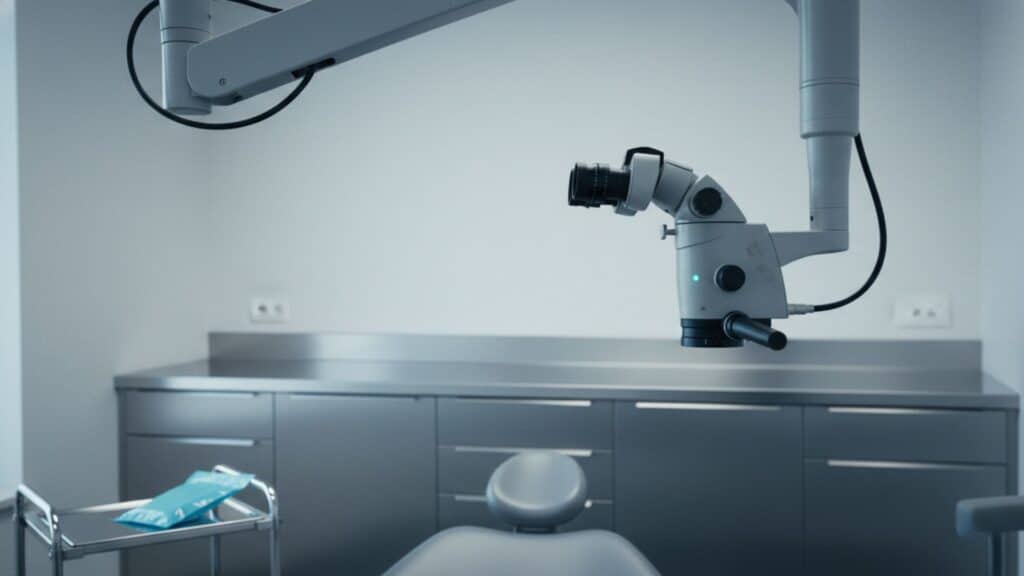
A dental (operating) microscope is a magnifying device with coaxial illumination that enlarges the working field 6× to 25× and lets the dentist see tooth detail invisible to the naked eye or to loupe glasses. In endodontics it reveals the fourth canal in upper molars; in retreatment it locates broken instruments; in aesthetic work it enables precision down to tenths of a millimetre.
Compared with the loupe glasses dentists commonly wear, the differences are clear. Loupes magnify only 2.5× to 4.5×, with a limited field of view and a fixed focal distance. An operating microscope offers 6× to 25× magnification, a wide field of view, smooth focusing, integrated coaxial illumination (LED or xenon) and the option of photo and video documentation.
In effect, the dentist sees the inside of a root canal as if from a few centimetres away, not tens.
How a microscope improves treatment success
Where, specifically, does the microscope shift the results?
Endodontics of upper molars. Vertucci (Endodontic Topics, 2005) showed that the fourth canal (MB2) is present in 78 % of upper first molars — yet without a microscope it is found in only 30–35 % of cases. A missed MB2 is the most common reason endodontic treatment fails.
Retreatment. A re-treated canal contains old filling material and often a wall perforation. Without a microscope, these obstacles are hard to locate and remove safely.
Detection of microfractures. A vertical root fracture is often visible only at around 16× magnification. Without a microscope it is diagnosed only after an unnecessary retreatment.
Conservative dentistry. Locating interproximal decay, checking filling margins, removing old composite without damaging healthy tissue — across all of this, the microscope raises both quality and success.
What is the microscope used for?
The microscope adds real value to almost every procedure we perform:
- Retreatment — always. Without a microscope, retreatment success drops by 15 to 25 percentage points.
- Endodontics of upper molars — because of the MB2 canal.
- Patients with anatomical anomalies — fused roots, calcified canals.
- Diagnosis of root microfractures.
- Aesthetic dentistry — veneers and anterior composites, where every tenth of a millimetre decides the result.
- Younger patients — preserving the most healthy tooth structure during preparation.
Frequently asked questions
Is a microscope at the dental clinic just marketing?
No — if the dentist is certified in microscopic endodontics and uses it routinely. If the microscope sits “in the corner for the photos”, then yes. Ask for concrete success-rate data and for the option of documenting the procedure.
Does treatment under a microscope hurt more?
No. Pain depends on the local anaesthetic and how gentle the procedure is. A microscope often allows a more conservative approach, because the dentist sees exactly what they are doing — and often removes less healthy tissue.
Does treatment under a microscope take longer?
Slightly, yes — typically 15 to 25 % longer. That extra time reflects more precise work. In practice it means 15–30 extra minutes for a root canal. The patient gets it back as a higher success rate and a lower risk of needing the treatment redone.
Conclusion
At m2stoma, we use a microscope in every procedure that calls for one. Each treatment is documented before, during and after, so you can see exactly what was done and how. Planning a retreatment or a more complex endodontic procedure? Call us, or book online.